Hyaluronic Acid Bioink

$99,00$280,00

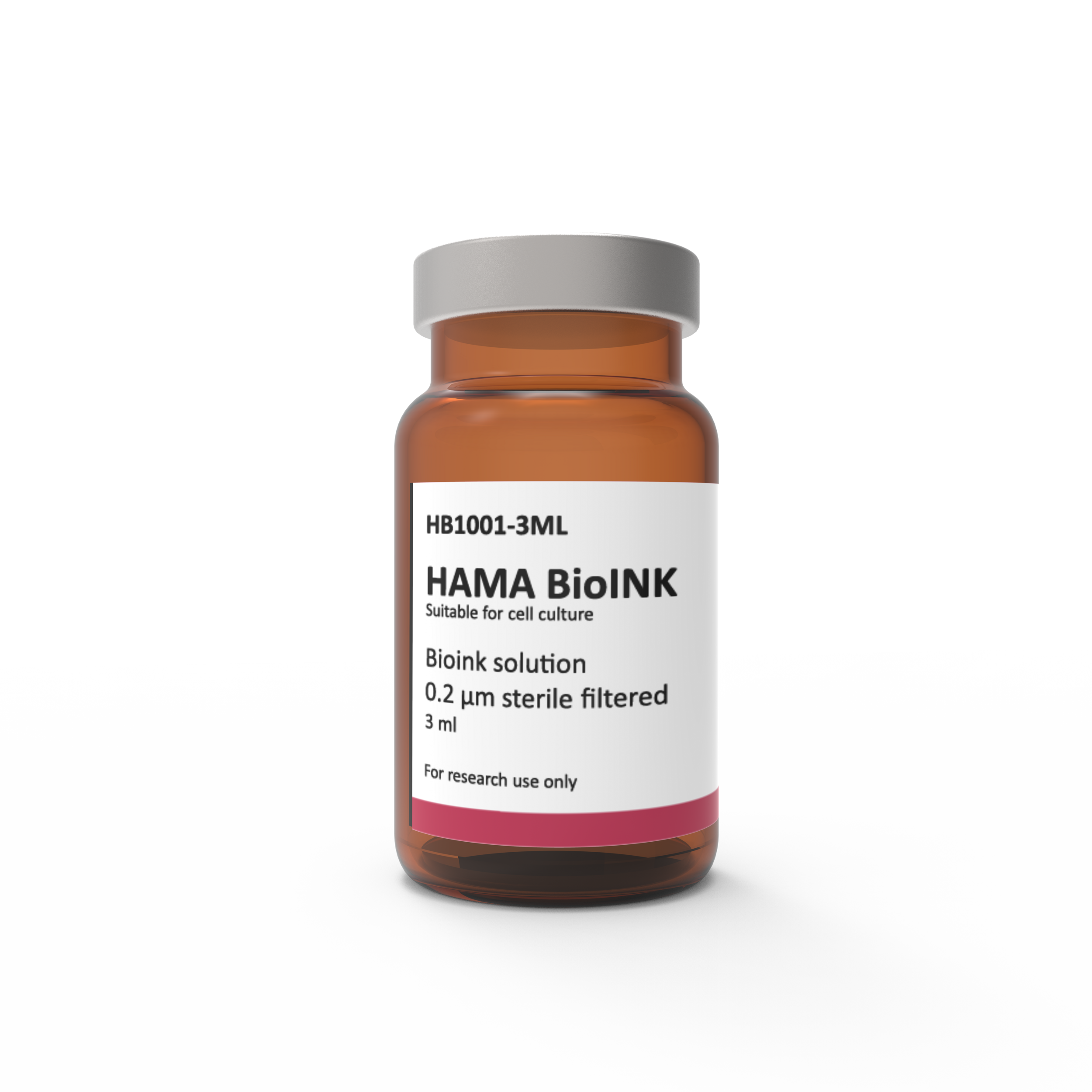
Adbioink offers methacrylated hyaluronic acid bioink. Hyaluronic acid (HA) [α-1,4-D-glucuronic acid-β-1,3-N-acetyl-D glucosamine]n, is a naturally occurring high molecular weight hydrophilic glycosaminoglycan. It is an abundant polymer in the extracellular matrix, where it provides mechanical support. Hyaluronan also provides to tissue hydrodynamics, movement and proliferation of cells, and participates in a number of cell surface receptor interactions. It is a cell-friendly material and oftenly used natural polymer for cell scaffolding.

The chemical structure of hyaluronic acid allows (primary) hydroxyl groups to be easily replaced by esterification. Modification with methacrylate groups followed by the addition of a photoinitiator leads to polymerization upon UV exposure, resulting in network formation. Methacrylated hyaluronic acid (HaMA) is more resistant to degradation and more rigid than non-derived hyaluronic acid hydrogels, while retaining its biocompatibility. Methacrylation increases the final mechanical strength of hyaluronic acid and this results in hyaluronic acid-based hydrogels that can be 3D bioprinted. These hydrogels shows good primary cell survival and superb spontaneous osteogenic differentiation in vitro. HaMA-based hydrogels with intrinsic osteogenicity are promising scaffold materials for application in 3D printed, tissue engineered bone replacements.

Description

Synonyms HAMA Bioink, Hyaluronan Bioink, Hyaluronic Acid Hydrogel Bioink, Hyaluronic Acid Bioink, Hyaluronic Acid Methacrylate Bioink, Methacrylated Hyaluronic Acid Bioink
Sterility Sterile
Endotoxin level <50 EU/mL
Cell viability ≥85% fibroblast cells, mesenchymal stem cells and osteoblast cells
pH 6.0-8.0
Form Gel

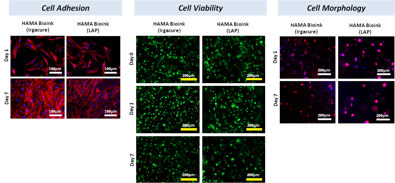
Hyaluronic acid methacrylate bioinks developed by AdBioInk Biosystem Corp. are prepared by using two different photoinitiators, Irgacure2959 and LAP. Cell adhesion and cell encapsulation experiments were performed against NIH-3T3 cell line. Cell viability of Hyaluronic acid bioinks has been determined by using Live/Dead kit. Results showed that cell-laden hyaluronic acid bioinks support cell viability (more than 95%). In Dapi-Actin staining images where cell morphology was examined, bioinks prepared with Irgacure and LAP supported cell adhesion and proliferation for 7 days.
Additional information

Volume

3ml, 3x3ml

Photoinitiator type

Irgacure 2959, LAP

Reviews

There are no reviews yet.

Be the first to review “Hyaluronic Acid Bioink”

Your email address will not be published. Required fields are marked *