GelMA Bioink

$99,00$280,00

GelMA (gelatin methacrylate) is a hydrolysis product of collagen derivative containing methacrylate groups. GelMA contains arginine-glycine-aspartic acid (RGD) sequences that promote cell attachment that are also suitable for cell remodeling. GelMA enables the hydrogel microfabrication to create unique patterns, morphologies, and 3D-structures, providing ideal platforms to control cellular behaviors, to study cell–biomaterial interactions, and to engineer tissues. The excellent biocompatibility of GelMA hydrogels makes them suitable as tissue scaffolds that mimic native ECM. Their compressive modulus are tuned by varying the degree of methacrylation degree, making it suitable for a wide range of biomedical applications.

Furthermore, GelMA could be a good candidate for bioink applications. 3D printed GelMA scaffolds have excellent cell viability and scaffold stability. The combination of nanotechnology and GelMA hydrogels will bring promising benefits in tissue engineering.

Easy to create personalized bioinks by following our adbioink protocols.

Documents

Description

Synonyms GelMA Bioink, GelMA Lyophilizate Bioink, Methacrylated Gelatin Bioink, Gelatin Methacrylate Bioink, Gelatin Methacryloyl Bioink
Sterility Sterile
Endotoxin level <50 EU/mL
Cell viability ≥85% fibroblast, osteoblast and mesenchymal stem cells
pH 7.3 – 7.4
Degree of methacrylation >80% (GelMA High)

50-60% (GelMA Middle)

Form Gel

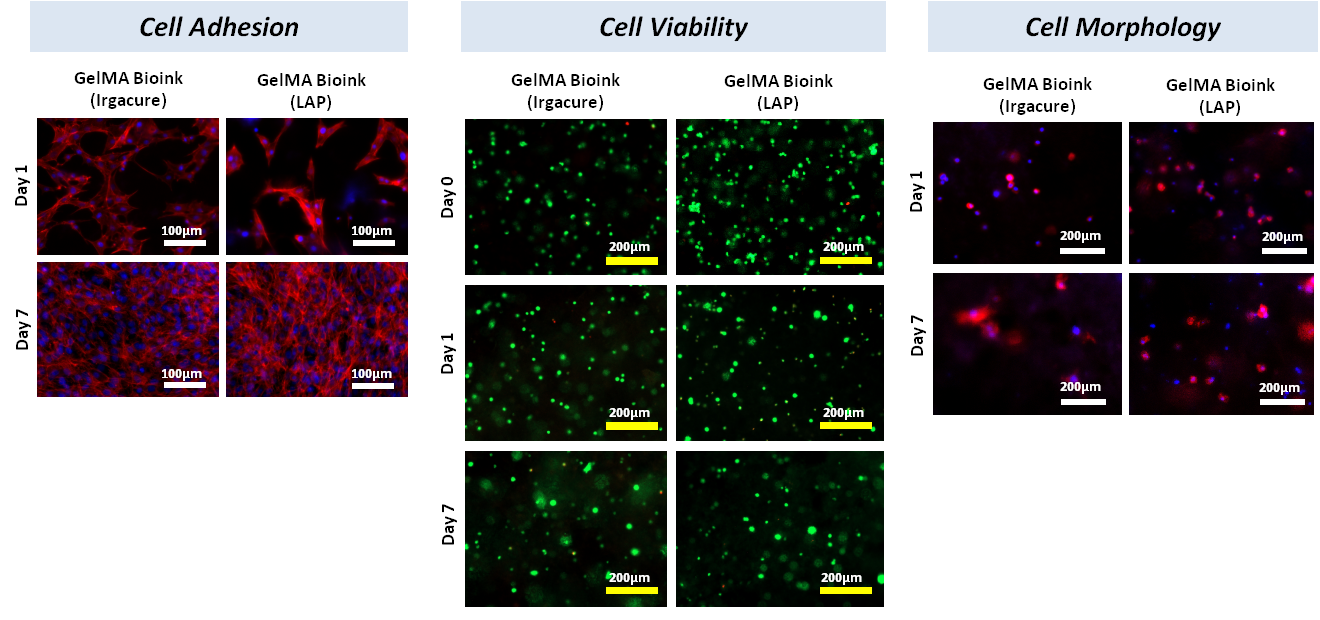
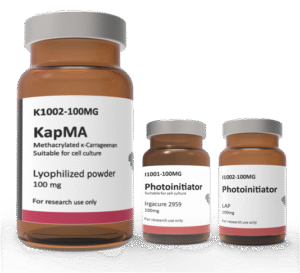
Gelatin methacrylate (GelMA) bioinks developed by AdBioInk Biosystem Corp. are prepared by using two different photoinitiators, Irgacure2959 and LAP. Cell adhesion and cell encapsulation experiments were performed against NIH-3T3 cell line. In cell viability assays, Live/Dead kit was used. It was observed that the cell viabilities in UV or visible light crosslinked and cell-encapsulated GelMA bioinks were over 95%.

Cell morphologies in and onto the crosslinked-GelMA bioinks were analyzed by DAPI-ACTIN staining. Cells seeded on GelMA bioinks took their cell morphology on day 1 and proliferated by day 7, showing over 90% confluence on bioink. Encapsulated cells appear to begin to show cytoplasmic protrusions at day 7.

Additional information

Volume

3ml, 3x3ml

Methacrylation degree

>80% (Gelma High), 50-60% (GelMA Middle)

Photoinitiator type

Irgacure 2959, LAP

Reviews

There are no reviews yet.

Be the first to review “GelMA Bioink”

Your email address will not be published. Required fields are marked *