
Description
| Synonyms | HAMA Bioink, Hyaluronan Bioink, Hyaluronic Acid Hydrogel Bioink, Hyaluronic Acid Bioink, Hyaluronic Acid Methacrylate Bioink, Methacrylated Hyaluronic Acid Bioink |
| Sterility | Sterile |
| Endotoxin level | <50 EU/mL |
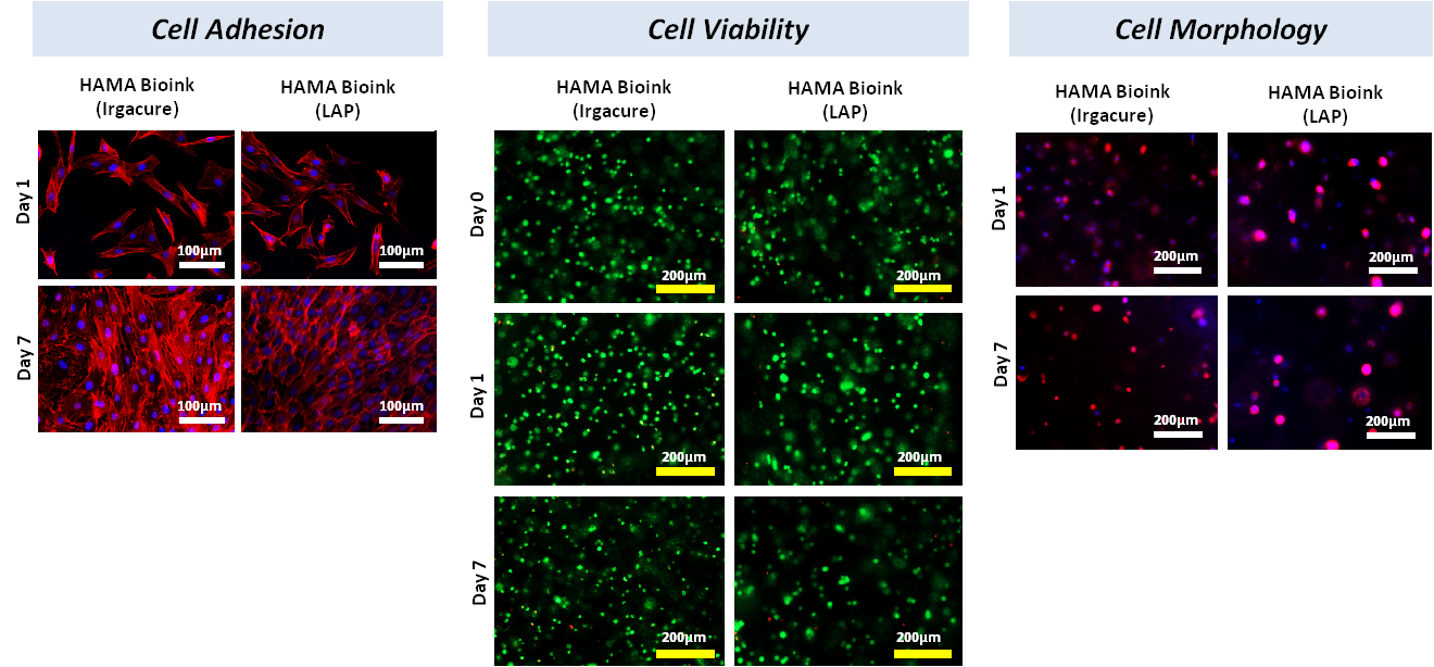
| Cell viability | ≥85% fibroblast cells, mesenchymal stem cells and osteoblast cells |
| pH | 6.0-8.0 |
| Form | Gel |
Hyaluronic acid methacrylate bioinks developed by AdBioInk Biosystem Corp. are prepared by using two different photoinitiators, Irgacure2959 and LAP. Cell adhesion and cell encapsulation experiments were performed against NIH-3T3 cell line. Cell viability of Hyaluronic acid bioinks has been determined by using Live/Dead kit. Results showed that cell-laden hyaluronic acid bioinks support cell viability (more than 95%). In Dapi-Actin staining images where cell morphology was examined, bioinks prepared with Irgacure and LAP supported cell adhesion and proliferation for 7 days.
Reviews
There are no reviews yet.